
Catheter ablation is one of the most effective treatments for most forms of paroxysmal supraventricular tachycardia (SVT).
This minimally invasive electrophysiology procedure precisely identifies the electrical mechanism responsible for the rapid heart rhythm. Energy is then applied to a very small area of heart tissue to eliminate the arrhythmia circuit or focus.
Medication usually reduces or prevents episodes without removing their underlying cause. In many forms of SVT, ablation can offer a definitive treatment, with very high success rates.
What you need to know
Open the topics you would like to read. The full guide is available in expandable sections.
01What is supraventricular tachycardia?+
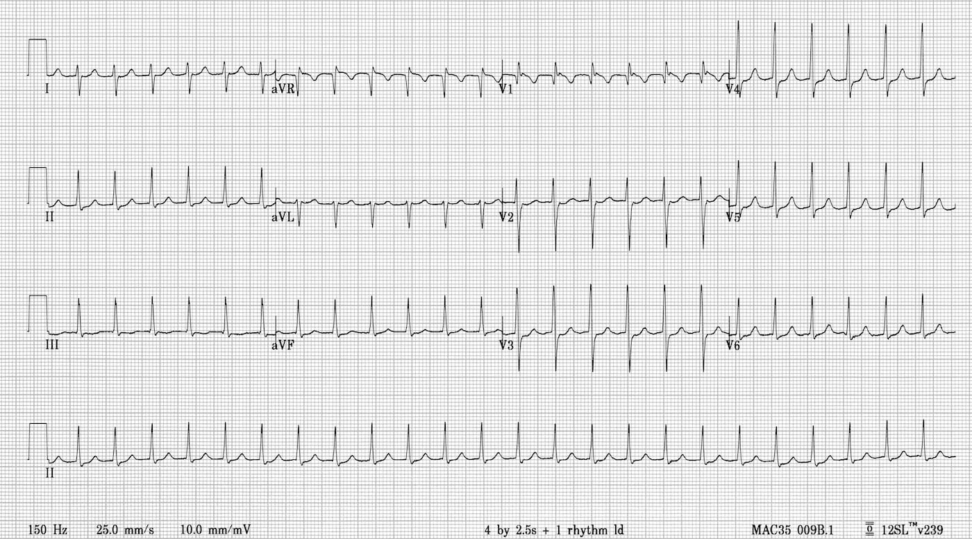
Supraventricular tachycardia (SVT) describes a group of arrhythmias originating in the atria or involving electrical circuits in the region between the atria and ventricles.
Episodes typically involve:
- a rapid heartbeat that starts and stops suddenly
- pronounced palpitations
- chest discomfort or pressure
- breathlessness
- dizziness or weakness
- near-fainting or, less commonly, fainting.
The heart rate during an episode is often between 150 and 220 beats per minute, although it may be lower or higher.
Most supraventricular tachycardias are not immediately life-threatening, but can significantly affect daily life and quality of life. When very frequent or prolonged, some forms can cause tachycardia-induced cardiomyopathy: weakening of heart function caused by a persistent rapid rhythm.
02Which types of SVT can be treated with ablation?+
The ablation technique depends on the precise electrical mechanism of the arrhythmia.
1. Atrioventricular Nodal Re-entrant Tachycardia – AVNRT
AVNRT (atrioventricular nodal re-entrant tachycardia) is one of the most common forms of paroxysmal SVT.
It results from a small electrical re-entry circuit around the atrioventricular node, usually involving two conduction pathways with different properties: a “fast” pathway and a “slow” pathway.
Ablation aims at slow pathway modification, preventing the tachycardia circuit from forming while preserving normal atrioventricular node function.
2. Atrioventricular Re-entrant Tachycardia – AVRT and Wolff-Parkinson-White Syndrome
In AVRT (atrioventricular re-entrant tachycardia), an additional electrical connection between the atria and ventricles is present, called an accessory pathway.
This pathway can participate in an electrical re-entry circuit and cause episodes of tachycardia.
When the accessory pathway conducts electrical impulses from the atria to the ventricles, it may produce characteristic pre-excitation on the ECG. When pre-excitation occurs together with clinical arrhythmias, the term Wolff-Parkinson-White (WPW) syndrome is used.
There are also concealed accessory pathways, which are not visible on the ECG during sinus rhythm but can participate in AVRT.
During the procedure, the precise location of the accessory pathway is identified and ablated to interrupt the abnormal electrical connection.
In selected patients with pre-excitation, an electrophysiology study and potentially ablation may be considered even without previous symptomatic tachycardia, if high-risk features or particular occupational requirements are present.
3. Focal Atrial Tachycardia
In focal atrial tachycardia, a particular area of the right or left atrium generates abnormally rapid electrical impulses.
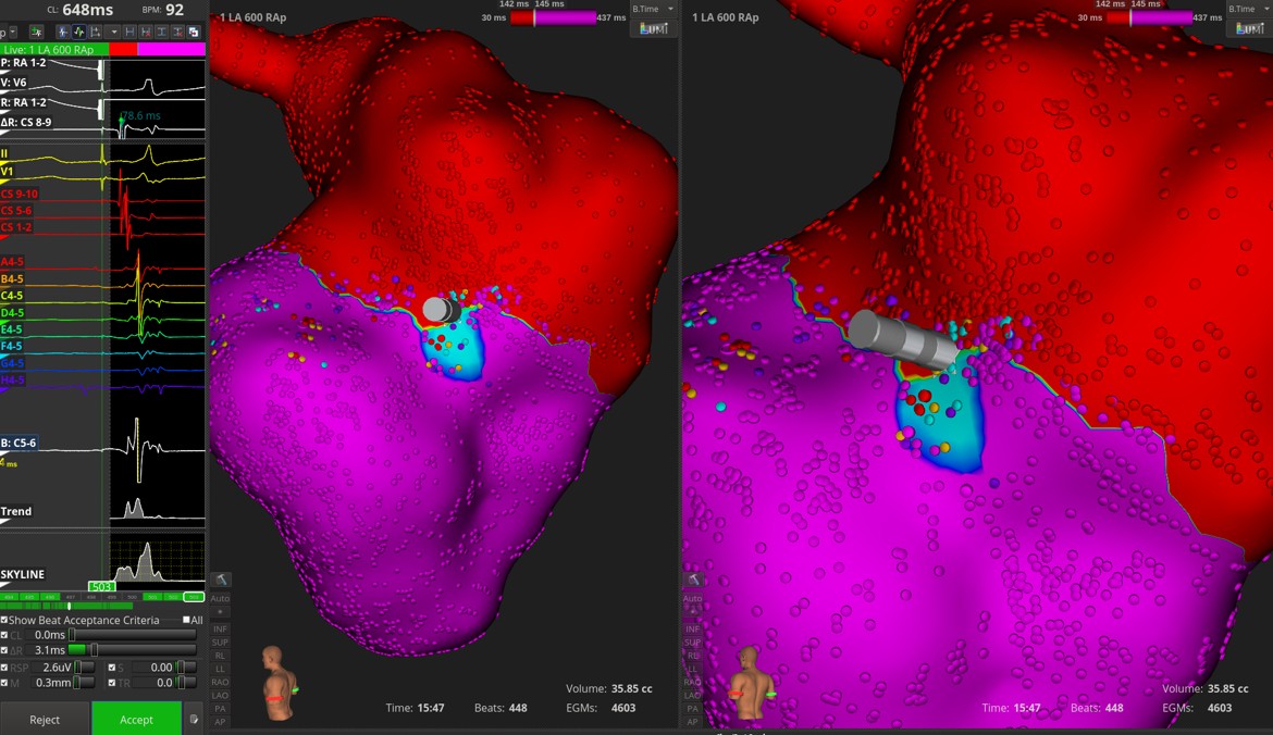
The precise location of this focus varies considerably between patients. Electrical mapping and, when required, three-dimensional electroanatomical mapping are used to locate it.
Once the site of origin is identified, ablation energy is applied to eliminate the abnormal focus.
03When is ablation recommended?+
Ablation may be the preferred treatment when there are:
- recurrent symptomatic episodes of SVT
- episodes that significantly affect daily life, work or exercise
- frequent emergency department visits
- a repeated need for adenosine or other emergency treatment
- significant haemodynamic compromise, near-fainting or fainting during tachycardia
- very frequent or almost continuous tachycardia
- tachycardia-induced cardiomyopathy
- medication that is ineffective or causes side effects
- a wish to avoid long-term antiarrhythmic treatment
- AVRT or WPW syndrome
- an accessory pathway with high-risk electrophysiological characteristics.
For AVNRT and AVRT, ablation is an established treatment and may be offered to suitable patients as a definitive alternative to long-term medication.
The choice is individualised according to the arrhythmia type, frequency and severity of episodes, age, coexisting cardiac conditions and patient preferences.
04Does the tachycardia need to have been recorded beforehand?+
Recording the arrhythmia on an ECG, Holter monitor or another recording device is particularly helpful, as it can provide important clues to its likely mechanism.
However, patients with a highly characteristic history of paroxysmal tachycardia may undergo an electrophysiology study even if a full episode has not previously been recorded.
05Preparing for the procedure+
A cardiology and arrhythmia assessment is performed before ablation. Depending on the case, this may require:
- an ECG
- an echocardiogram
- Holter monitoring or another arrhythmia recording
- blood tests
- kidney function and electrolyte tests
- a pregnancy test, when indicated
- additional imaging in more complex cases.
Medication
Some antiarrhythmic medicines, beta-blockers or other treatments affecting heart rate may need to be temporarily withheld to make the arrhythmia easier to induce and diagnose accurately.
Do not stop any medication without specific instructions from your electrophysiologist.
Patients taking anticoagulants or antiplatelet medicines will also receive specific instructions.
Fasting
You will receive instructions about eating and drinking based on the time of the procedure and the planned sedation or anaesthesia.
06What happens during the procedure?+
The procedure takes place in a specially equipped electrophysiology laboratory.
Venous access
After local anaesthesia and venous puncture, thin electrophysiology catheters are usually introduced through veins in the groin and advanced to the heart using X-ray guidance. Venous puncture is performed under ultrasound guidance.
No surgical incision in the chest is needed.
Electrophysiology study
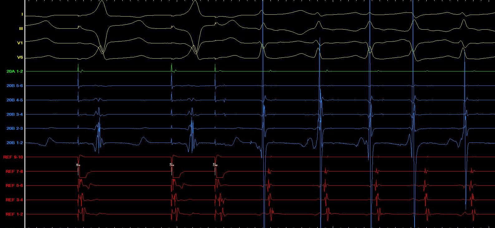
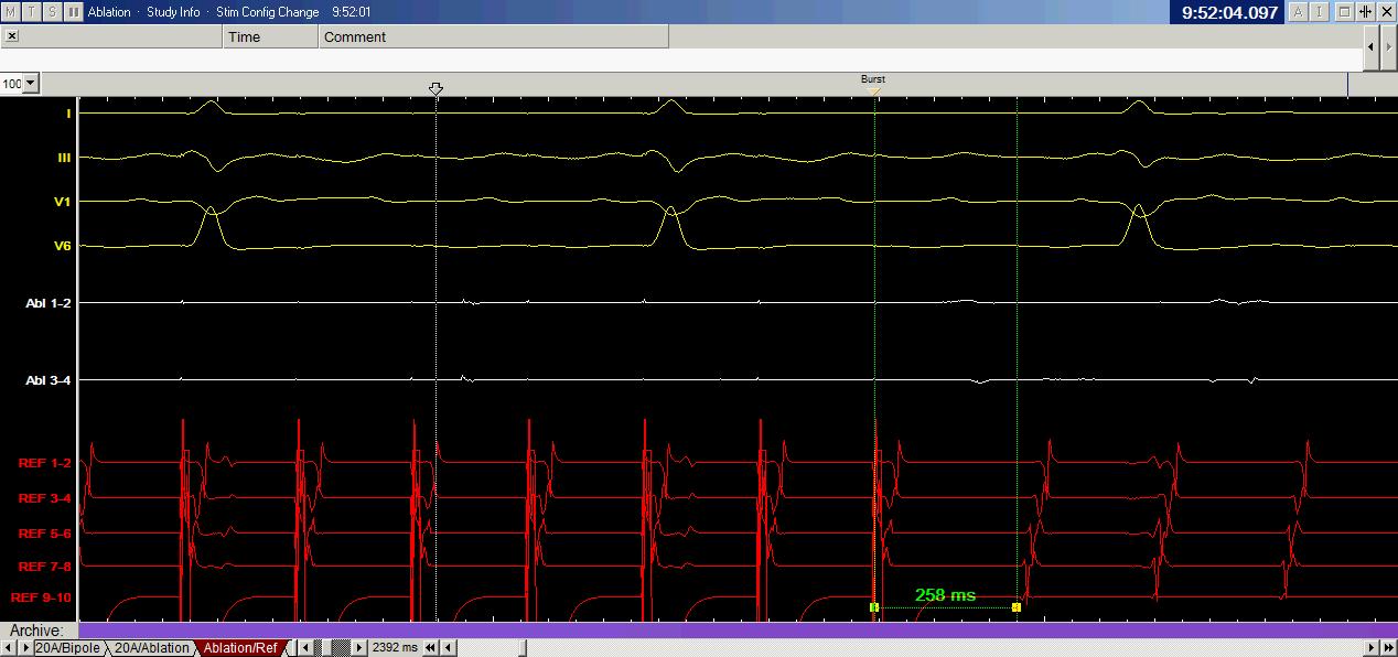
The catheters record electrical signals from different parts of the heart. Programmed electrical stimulation is used to induce the tachycardia in a controlled setting and identify its precise mechanism.
You may briefly feel the same rapid heartbeat as during your usual episodes. This is an expected part of the test and takes place under continuous monitoring.
In some cases, medicines such as isoprenaline are used to help induce the arrhythmia.
Mapping and ablation
Once the mechanism is established, the area involved in the arrhythmia is located precisely. Three-dimensional electroanatomical mapping systems are used in more complex cases.
Energy is then delivered through a specialised catheter at the appropriate site.
Electrophysiological testing is repeated after ablation to confirm that the intended result has been achieved and that the clinical tachycardia can no longer be induced.
07Which type of energy is used?+
Radiofrequency Ablation – RF
The most commonly used technique is radiofrequency (RF) ablation.
RF energy produces controlled local heating of a very small area of heart tissue, creating a small permanent lesion that prevents conduction of the abnormal electrical impulse.
It is widely used for:
- AVNRT
- AVRT and accessory pathways
- WPW
- focal atrial tachycardias.
Cryoablation
In selected cases, cryoablation may be used to cool tissue rather than heat it.
It can be particularly helpful when the ablation target lies close to the heart's normal conduction system, as it allows controlled and potentially reversible cooling before a permanent lesion is created.
It may be chosen for:
- some cases of AVNRT
- septal accessory pathways
- particular anatomical circumstances or younger patients.
The appropriate energy is selected according to the arrhythmia type and location, balancing effectiveness and safety for each patient.
08How long does the procedure take?+
A typical AVNRT or AVRT procedure can often be completed in approximately 1–2 hours, while more complex cases may take longer.
Duration depends on:
- the arrhythmia mechanism
- how easily it can be induced
- the location of the focus or accessory pathway
- whether access to the left side of the heart is needed
- the complexity of mapping.
09Is general anaesthesia needed?+
Most SVT ablation procedures can be performed with:
- local anaesthesia in the groin and
- light or moderate intravenous sedation.
Very deep sedation is often avoided because it can make the arrhythmia harder to induce.
Deeper sedation or general anaesthesia may be selected in particular or more complex cases.
10What if the tachycardia cannot be induced?+
In some cases, the clinical tachycardia cannot be induced during the electrophysiology study.
Further management depends on:
- previous ECG recordings
- findings from the electrophysiology study
- the most likely arrhythmia mechanism
- the safety of any proposed empirical ablation.
When there is sufficient electrophysiological evidence, ablation may be performed in selected patients even without inducing sustained tachycardia. If the mechanism cannot be established safely, the decision may be made not to ablate.
11What are the success rates?+
Success rates depend on the precise type and anatomical location of the arrhythmia.
AVNRT
Procedural success is usually around 95–98% or higher, with a low likelihood of recurrence.
AVRT / WPW
For most accessory pathways, success is above 95%, approaching 98–99% at many anatomical locations.
Pathways in more difficult locations may have slightly lower success rates or a higher chance of recurrence.
Focal atrial tachycardia
Success is also high, but depends more on the location of the focus, the mechanism and the underlying cardiac anatomy. In many patient series, acute success rates are approximately 85–95% or higher, depending on the type of tachycardia.
12Can the tachycardia return?+
Recurrence after successful AVNRT or AVRT ablation is relatively uncommon.
If the same arrhythmia returns, another electrophysiology study and repeat ablation can be performed, usually with a high likelihood of success.
Complex or scar-related atrial tachycardias may be more likely to recur than AVNRT or AVRT.
13What are the risks?+
Ablation of most straightforward SVTs is considered a low-risk procedure, particularly when performed by a specialist electrophysiology team.
The precise risks depend on the arrhythmia type, the anatomical location of the target and whether access to the left side of the heart is required.
Groin haematoma or bleeding
Minor bruising or tenderness at the catheter entry site is relatively common and usually settles without specific treatment.
A larger haematoma or another vascular complication is much less common.
Vascular injury
Rarely, a vein or artery used for catheter access may be injured.
Pericardial effusion or cardiac tamponade
Very rarely, injury to the heart wall can cause blood to collect in the pericardial space.
This risk is particularly low in straightforward right-sided SVT procedures.
Atrioventricular block
This is the most important specific risk of AVNRT ablation and ablation of certain accessory pathways close to the atrioventricular node.
If permanent complete atrioventricular block occurs, pacemaker implantation may be needed.
For typical AVNRT treated with modern techniques, the risk of permanent AV block is very low, below 0.5%.
The risk may be higher for some septal accessory pathways because of their proximity to the normal conduction system.
Thromboembolism or stroke
The risk is very low for straightforward right-sided SVT ablation and increases slightly when access to the left side of the heart is required.
Coronary injury
Extremely rarely, a coronary artery may be injured when the ablation site is very close to it.
Complications of sedation or medication
Rare complications include an allergic reaction, low blood pressure, a slow heart rate or respiratory depression. You are therefore monitored continuously throughout the procedure.
14What happens afterwards?+
After the catheters are removed, pressure is applied to the venous access sites and you remain under observation for a period of time.
You will usually need to lie flat for a few hours to reduce the risk of bleeding from the groin.
Most straightforward procedures involve a short hospital stay. You can usually return home on the same or following day, depending on the timing and type of procedure and your clinical condition.
15What might I feel in the first few days?+
After the procedure, you may notice:
- mild tenderness or bruising in the groin
- occasional ectopic beats
- brief palpitations.
These symptoms do not necessarily mean that the arrhythmia has returned.
If a prolonged episode resembling your previous tachycardia occurs, it is helpful to obtain an ECG recording if possible.
16Returning to everyday activities+
Most patients return quickly to everyday activities.
For the first few days, you will usually be advised to avoid:
- strenuous exercise
- heavy lifting
- activities that place significant pressure on the groin.
The timing of return to work, driving and strenuous exercise is individualised according to the procedure and recovery.
17Will I need medication after ablation?+
After successful AVNRT or AVRT ablation, long-term antiarrhythmic treatment for that particular arrhythmia is usually no longer needed.
However, no medication should be stopped without medical advice, as it may also have been prescribed for another reason.
Some left-sided procedures may require temporary antithrombotic or anticoagulant treatment, depending on the technique and the patient's characteristics.
18When should I seek urgent medical attention?+
After discharge, seek immediate medical assessment if you develop:
- groin bleeding that does not stop with pressure
- rapidly increasing swelling or a haematoma
- severe or worsening chest pain
- significant breathlessness
- fainting
- persistent tachycardia
- weakness, numbness or difficulty speaking
- a high fever or other signs of infection.
19Ablation or medication?+
The choice depends on the arrhythmia type, frequency of episodes, symptom severity and patient preferences.
Medication may be appropriate when episodes are infrequent and well tolerated, or when the patient does not wish to undergo an invasive procedure.
However, medicines usually do not eliminate the electrical substrate of the arrhythmia. Particularly for AVNRT and AVRT, ablation offers the possibility of definitive treatment with very high success rates, without the need for long-term antiarrhythmic medication in most patients.
20Individualised treatment+
There is no single form of “SVT ablation”. The procedure is tailored to the exact mechanism and anatomical location of the arrhythmia.
The electrophysiology study and ablation are usually performed during the same procedure: the mechanism is established first, followed by ablation if a suitable and safe target is identified.
With modern electrophysiology and mapping techniques, most forms of paroxysmal SVT can be treated with very high success rates and a low risk of complications.
This page provides general information. The indication, technique, likelihood of success and specific risks of ablation are individually assessed by your doctor.