
Ablation uses three-dimensional mapping to locate the arrhythmogenic focus and deliver targeted energy, aiming to reduce symptoms and PVC burden.
What you need to know
Open the topics you would like to read. The complete guide is available in expandable sections.
01What are premature ventricular contractions (PVCs)?+
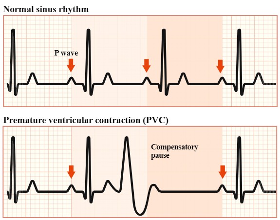
Premature ventricular contractions (PVCs) are early heartbeats originating in the ventricles rather than the normal electrical system that sets the heart's rhythm.
They are very common and occur occasionally in many people without necessarily indicating heart disease. However, when very frequent or symptomatic, they can significantly affect quality of life. In some patients, a high number of ectopic beats over a prolonged period can lead to impaired heart function (PVC-induced cardiomyopathy).
Common symptoms include:
- a fluttering sensation or forceful heartbeats
- a feeling that the heart “misses” a beat
- a pause followed by a strong heartbeat
- a racing heart
- dizziness or unsteadiness
- breathlessness
- fatigue
- reduced exercise tolerance
In many patients, PVCs are completely asymptomatic and are found incidentally on an ECG or Holter recording.
02Where do PVCs originate?+
PVCs can arise from different areas of the right or left ventricle. Their appearance on a 12-lead ECG provides important information about their likely origin.
Common sites include:
- the right ventricular outflow tract (RVOT)
- the left ventricular outflow tract (LVOT)
- the sinuses of Valsalva / aortic root
- the Purkinje system and left ventricular fascicles
- the papillary muscles
- the mitral or tricuspid annulus
- the LV summit
- less commonly, intramural or epicardial regions.
The precise location is important because it affects the ablation technique, likelihood of success and potential complications.
03Are PVCs dangerous?+
In most patients with a structurally normal heart, idiopathic PVCs have a benign prognosis.
An individual assessment is nevertheless needed to exclude underlying heart disease and determine the frequency and origin of the ectopic beats.
The PVC burden is particularly important: this is the percentage of all heartbeats that are PVCs during a Holter recording.
In some patients, a high PVC burden can gradually cause the left ventricle to dilate and contract less effectively. This is called PVC-induced cardiomyopathy and may be reversible when the PVCs are effectively suppressed.
04What is PVC ablation?+
PVC catheter ablation is a minimally invasive electrophysiology procedure that aims to:
- identify precisely the area of the heart where the ectopic beats originate and
- eliminate that arrhythmogenic focus.
Specialised thin catheters are introduced into the heart through blood vessels, without a surgical incision in the chest.
05When is ablation recommended?+
The decision is individualised, taking account of:
- PVC frequency
- symptom severity
- the site of origin
- left ventricular function
- whether underlying heart disease is present
- the effectiveness and tolerability of medication
- patient preferences.
Ablation may be particularly indicated for:
Symptomatic PVCs
When ectopic beats cause significant symptoms and affect daily life or quality of life.
Very frequent PVCs
When the PVC burden is high and there is concern about its possible effect on heart function.
PVC-induced cardiomyopathy
When the ejection fraction is reduced and frequent PVCs are considered the cause or an important contributing factor.
Effective elimination of PVCs may then lead to substantial improvement or even restoration of left ventricular function.
Ineffective or poorly tolerated medication
When medicines do not adequately control the arrhythmia or cause side effects.
Specific idiopathic PVCs
For symptomatic idiopathic PVCs originating from the RVOT or left fascicles, ablation is a first-line treatment according to contemporary European guidelines.
06Which tests are needed before ablation?+
Pre-procedural assessment is tailored to each patient and may include:
- a 12-lead ECG
- Holter monitoring to assess PVC burden and morphology
- an echocardiogram
- blood tests
- an exercise test where appropriate
- cardiac MRI in selected patients.
MRI is particularly useful when underlying cardiomyopathy or myocardial scar needs to be excluded.
Recording PVCs on a 12-lead ECG beforehand is especially helpful because their morphology can guide the electrophysiologist towards the likely site of origin.
07Preparing for the procedure+
You will receive specific instructions before the procedure.
Depending on your circumstances, you may be asked to temporarily stop certain antiarrhythmic medicines, so that PVCs are present during the procedure and can be mapped.
Do not stop any medicine without specific medical instructions.
Fasting for a specified number of hours is usually required. You will be told which medicines you can take on the day.
08What happens during ablation?+
1. Vascular access
After local anaesthesia, thin catheters are usually introduced through a vein in the groin and advanced towards the heart.
Depending on where the PVCs originate, access to the left ventricle may be required. This can be achieved through the aorta (retrograde aortic access) or, in appropriate cases, by transseptal puncture.
2. Mapping
The most important stage is locating the arrhythmogenic focus accurately.
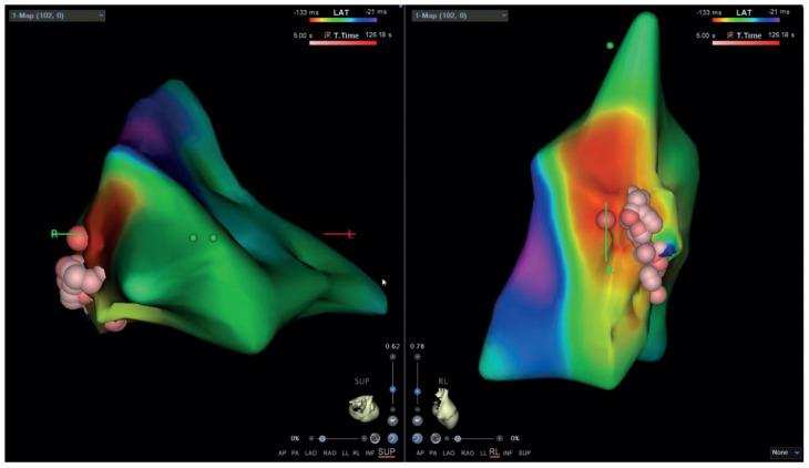
Advanced three-dimensional electroanatomical mapping systems create an electrical and anatomical map of the heart.
Mapping may use two main techniques:
Activation mapping
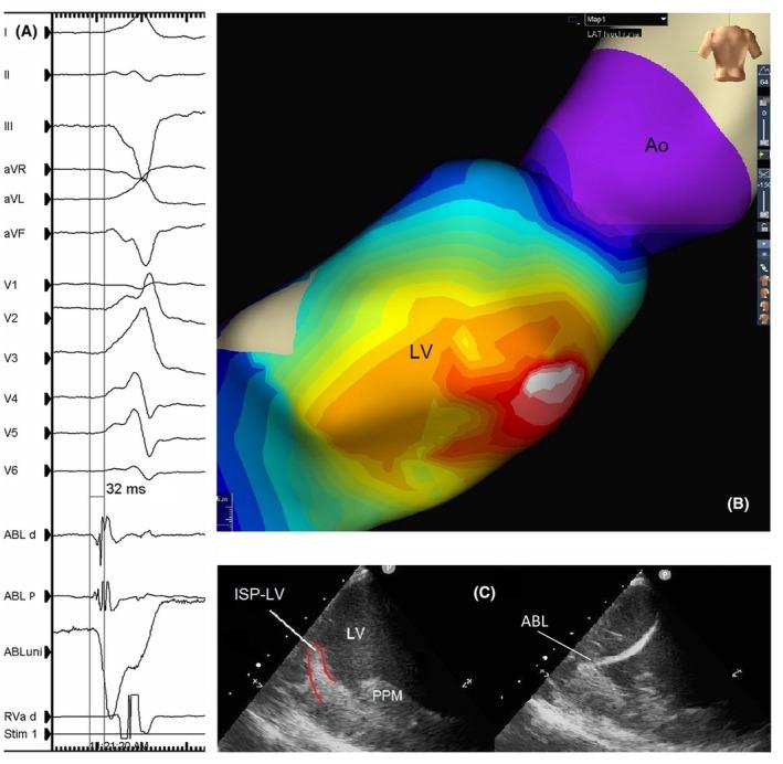
When PVCs occur frequently during the procedure, electrical activation is recorded at different points in the ventricle.
The aim is to identify the site where electrical activation precedes the onset of the PVC on the ECG by the greatest interval.
Pace mapping
Controlled electrical stimulation is delivered at different ventricular sites, and the resulting QRS morphology is compared with that of the spontaneous PVC.
The closer the match, the more likely the site is to be near the true focus.
09Why do PVCs need to be present on the day?+
Unlike many other ablation procedures, PVC ablation depends substantially on the ability to record and map ectopic beats during the procedure.
If PVCs are very infrequent that day, locating the focus accurately may be more difficult.
Special techniques or medicines may therefore be used to provoke PVCs or increase their frequency.
This is also why suppressing PVCs with certain antiarrhythmic medicines beforehand may be undesirable, and why medication may be temporarily adjusted under specific medical instructions.
10Which type of energy is used?+
Radiofrequency (RF) ablation
The most commonly used technique is radiofrequency ablation.
Controlled energy is delivered through a specialised catheter tip to the site where the arrhythmia originates. It creates a very small, localised thermal lesion in the arrhythmogenic tissue to eliminate the focus.
Modern catheters allow monitoring of parameters such as contact with the myocardium, power, temperature and duration of energy delivery.
Cryoablation
At selected anatomical sites, cryoablation may be used to cool tissue rather than heat it.
The choice depends on the arrhythmia's origin and its proximity to important anatomical structures.
11Is general anaesthesia needed?+
Ablation can be performed under local anaesthesia with sedation, although in the majority of cases it is performed under general anaesthesia.
Anaesthesia is planned carefully for PVC ablation because some anaesthetic agents or deep sedation may reduce PVC frequency and make mapping more difficult.
The approach is therefore tailored to the arrhythmia, the patient and the expected duration and complexity of the procedure.
12How long does the procedure take?+
Duration varies considerably according to:
- the site of origin
- PVC frequency during the procedure
- whether left ventricular mapping is needed
- whether more than one PVC morphology is present
- anatomical complexity.
A relatively straightforward focus may be treated more quickly, while complex cases can require several hours of mapping and ablation.
13What happens immediately afterwards?+
The catheters are removed and pressure or a dedicated haemostasis system is applied to the vascular access site.
You will remain under observation and may need to lie flat for several hours.
Depending on the procedure and your clinical condition, you may be discharged the following day.
Before discharge, you will receive individual instructions about:
- medication
- care of the puncture site
- physical activity
- returning to work
- restarting exercise
- scheduled cardiology follow-up.
14What should I expect in the first few days?+
Mild discomfort or minor bruising in the groin is relatively common.
Some patients also notice occasional ectopic beats afterwards. A few PVCs immediately after ablation do not necessarily mean that the procedure has failed.
Long-term effectiveness is usually assessed through clinical review, an ECG and repeat Holter monitoring.
In patients with PVC-induced cardiomyopathy, left ventricular function is also reassessed, as the ejection fraction may improve gradually after successful PVC suppression.
15What is the success rate?+
The likelihood of success depends considerably on whether ventricular tachycardia is idiopathic or associated with structural heart disease. In structural heart disease, ablation is usually more complex because the arrhythmia may arise from scar tissue and involve more than one re-entry circuit.
VT ablation in ischaemic cardiomyopathy
In ischaemic cardiomyopathy, ventricular tachycardia is usually associated with scar following a myocardial infarction. Scar may be endocardial, epicardial or deeper within the myocardium, creating narrow electrical channels that sustain tachycardia.
Ablation aims to:
- interrupt re-entry circuits within or around the scar
- reduce VT recurrences
- reduce repeated ICD shocks
- reduce hospital admissions and the impact of electrical storms.
At experienced centres, acute elimination or non-inducibility of clinically important VTs is often achieved in approximately 80% of patients. This depends on scar extent and location, haemodynamic tolerance of VT and whether an endocardial, epicardial or combined approach is required.
Long-term freedom from VT recurrence is lower than immediate technical success. In many series, approximately 70–80% remain free of significant recurrence over the following months to one year. Recurrence risk increases with extensive scar, severe left ventricular dysfunction or multiple VT morphologies.
Ablation does not eliminate the risk of sudden cardiac death and usually does not replace an implantable cardioverter defibrillator (ICD) when one is indicated. Its principal aim is to substantially reduce episodes and ICD therapies, rather than remove the need for protection against future dangerous arrhythmias.
VT ablation in non-ischaemic cardiomyopathy
In non-ischaemic cardiomyopathy, the scar and arrhythmogenic substrate may have a different distribution and are often more diffuse, intramural or epicardial. VT may be associated with dilated cardiomyopathy, myocarditis, sarcoidosis, arrhythmogenic cardiomyopathy or other genetic and inflammatory conditions.
Ablation is often more technically demanding than in ischaemic cardiomyopathy because:
- the substrate may lie on the outer surface of the heart or deep within the myocardium
- multiple, separate scars may be present
- VT may be haemodynamically intolerable
- the arrhythmia may recur from a site that was initially inaccessible or not apparent
- in conditions such as sarcoidosis or myocarditis, ongoing inflammation may create new arrhythmogenic substrate.
Acute success in non-ischaemic cardiomyopathy is approximately 60–80%, but varies considerably with the cause and anatomical distribution of scar. Long-term freedom from recurrence is usually lower, approximately 40–60% over six to twelve months. Outcomes are better when scar is limited and localised, and more challenging when it is extensive, epicardial or multifocal.
Some patients with non-ischaemic cardiomyopathy require epicardial ablation, either initially or later, if the arrhythmogenic area cannot be adequately treated from the endocardium. The need for epicardial access depends on scar imaging, the ECG, electroanatomical mapping and the response to initial ablation.
What does “success” mean in VT associated with structural heart disease?
Success is not assessed solely by immediate elimination of tachycardia during the procedure. Assessment usually includes:
- non-inducibility of clinical VTs at the end of the procedure
- a substantial reduction in VT episodes afterwards
- fewer appropriate ICD therapies
- avoidance of electrical storms
- fewer hospital admissions
- improved quality of life.
Ablation may therefore be clinically successful even if it does not eliminate every possible VT, provided it substantially reduces dangerous episodes and ICD therapies.
Factors affecting the likelihood of success
Prognosis and recurrence risk depend on:
- the cause of the cardiomyopathy
- scar extent and location on cardiac MRI
- whether scar is endocardial, epicardial or intramural
- the number and morphology of VTs
- left ventricular function
- the presence of heart failure
- active inflammation
- haemodynamic tolerance of VT
- the need for epicardial or combined ablation
- the experience of the specialist centre.
Ablation for VT in structural heart disease is therefore primarily a treatment to reduce arrhythmia burden and dangerous recurrences. It does not treat the underlying cardiomyopathy or always remove the need for an ICD, medication or close follow-up.
For idiopathic VT, where there is no significant structural heart disease, success rates are usually higher and often exceed 80–90%, particularly when the arrhythmia arises from a well-localised, accessible focus. These estimates should not be applied to more complex VT associated with ischaemic or non-ischaemic cardiomyopathy.
The final prognosis is assessed individually, based on the cause of cardiomyopathy, scar imaging, the arrhythmogenic substrate and the experience of the treating team.
16What are the risks?+
PVC ablation generally has a high level of safety, particularly for idiopathic PVCs from common anatomical sites.
However, like any invasive cardiac procedure, it carries a small but real risk of complications.
Potential complications include:
Vascular complications
- groin haematoma
- bleeding
- pseudoaneurysm
- less commonly, vascular injury.
Cardiac complications
- myocardial perforation
- pericardial effusion
- cardiac tamponade, which may require emergency drainage.
Thromboembolic complications
Procedures in the left ventricle carry a small risk of clot formation and stroke or another embolic event. Appropriate anticoagulation is used during the procedure to reduce this risk.
Injury to the normal conduction system
If the focus lies close to the His bundle or the main conduction fascicles, there is a risk of impaired atrioventricular conduction.
Very rarely, significant conduction system injury may create a need for a permanent pacemaker.
Coronary artery injury
Some arrhythmogenic foci lie close to coronary arteries. Special precautions are taken in these cases, and coronary imaging may be needed before energy is applied.
Heart valve injury
This is rare but may occur with particular access routes or ablation sites.
Other rare complications
These include allergic reactions, complications of anaesthesia or medication, infection and, extremely rarely, a serious life-threatening complication.
Individual risk depends on the PVC origin, required access route, underlying heart disease and other medical conditions.
17Can PVCs return?+
Yes. Recurrence is possible even after successful ablation.
This may happen if:
- the original focus recovers electrical activity
- the true focus lies deeper within the myocardium
- more than one focus is present
- a new PVC morphology develops in the future.
Repeat ablation may be considered in selected cases.
18PVCs and cardiomyopathy+
The relationship between frequent PVCs and left ventricular function is particularly important.
Contemporary guidelines recognise that a PVC burden of approximately ≥10% may be associated with left ventricular dysfunction. Risk rises further at higher burdens, particularly above approximately 20%.
However, PVC burden is not the only risk factor, and not every patient with frequent PVCs develops cardiomyopathy.
When PVC-induced cardiomyopathy is suspected, ablation is a particularly effective treatment option and may lead to reverse remodelling and substantial recovery of heart function.
19Is ablation suitable for everyone with PVCs?+
No.
The presence of PVCs alone is not an indication for a procedure.
An asymptomatic patient with normal heart function and a low PVC burden may only need monitoring.
The decision is based on the overall clinical picture, not simply the number of ectopic beats.
Three key questions should therefore be answered before making a decision:
Where do the PVCs originate?
How frequent are they?
Do they cause symptoms or affect heart function?
The answers help determine the most appropriate treatment for each patient.
Individual assessment
PVC ablation is a specialist electrophysiology procedure. The strategy varies between patients and is determined by PVC morphology, anatomical origin and burden, together with heart structure and function.
Detailed assessment of the ECG, Holter recording and cardiac imaging helps establish whether treatment is needed and, when ablation is indicated, plan the safest and most effective approach.