
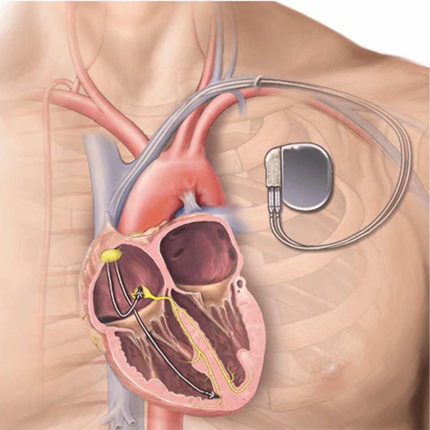
Pacemaker implantation is one of the most established treatments for patients whose heart rhythm is too slow or whose electrical conduction between the heart chambers is significantly impaired.
Modern pacing techniques have advanced considerably. Alongside conventional pacemakers, selected patients may benefit from cardiac resynchronisation therapy (CRT-P) or pacing of the heart's natural conduction system, such as left bundle branch area pacing (LBBAP).
The appropriate method is chosen individually according to the type of bradycardia or conduction disorder, heart function, ECG, expected need for ventricular pacing and the presence or absence of heart failure.
What is a pacemaker?
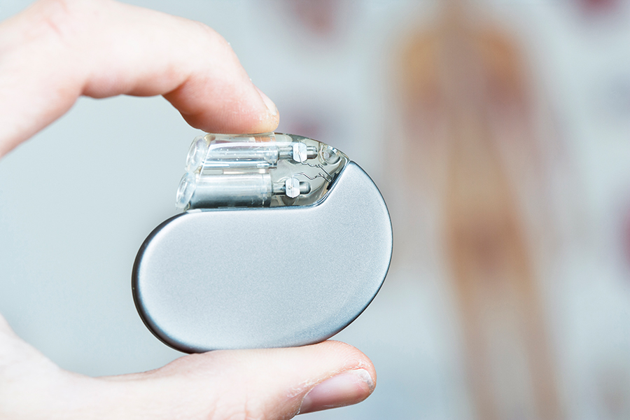
A pacemaker is a small electronic device that continuously monitors the heart rhythm. When the heart beats more slowly than intended, or electrical impulses are not conducted properly, it delivers small, painless impulses to stimulate the heart muscle and maintain an appropriate rhythm.
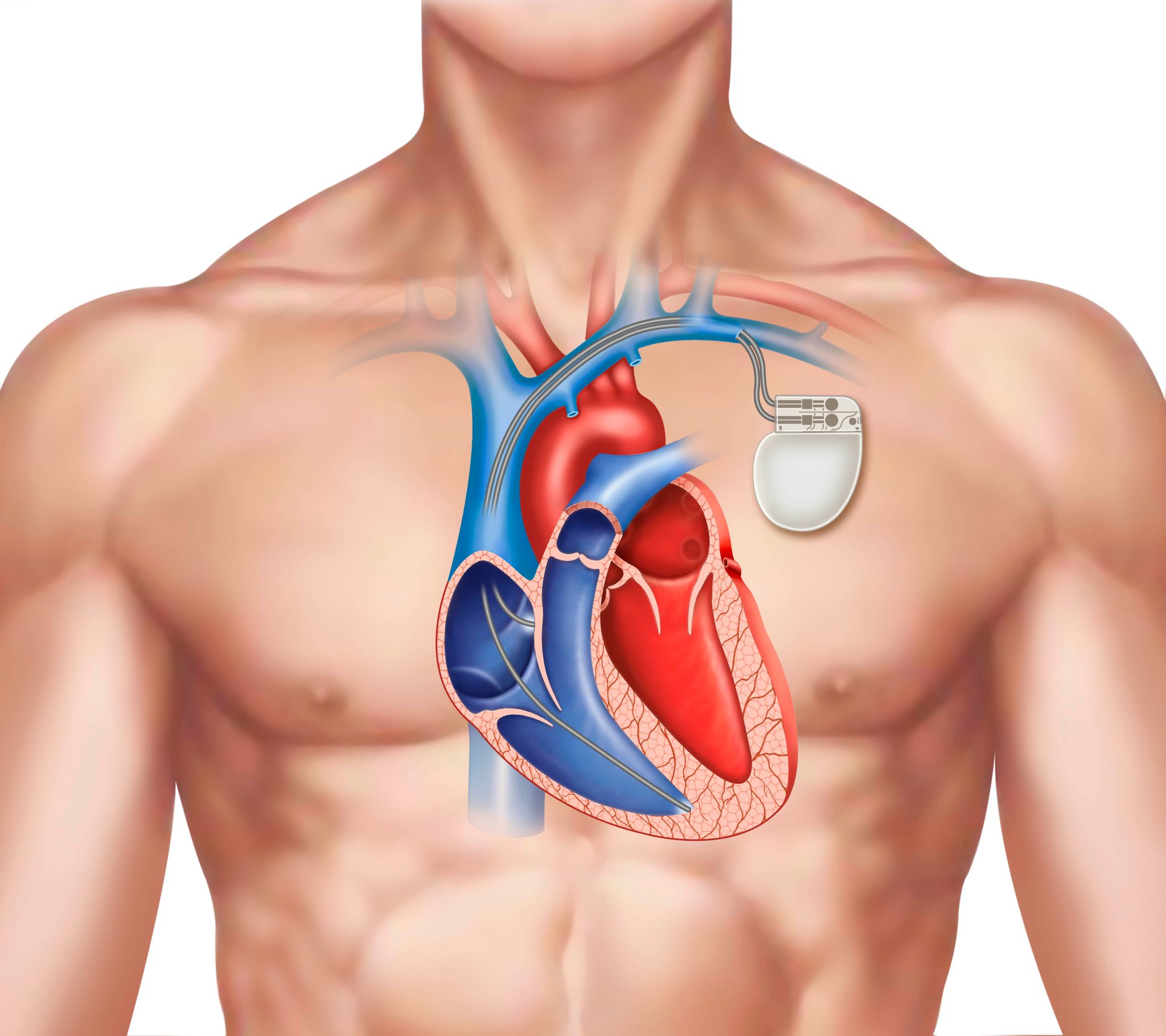
The system usually consists of:
- a generator containing the battery and electronic circuitry,
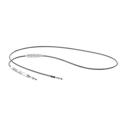
- and one or more leads introduced through a vein and positioned inside the heart.
Depending on the condition, a pacemaker with one, two or more leads may be used.
The pacemaker works only when needed. Some patients require pacing almost continuously, while others need it only occasionally.
When is a pacemaker needed?
Pacemaker implantation may be recommended for clinically significant bradycardia or a disorder of the heart's electrical system.
Common indications include:
Sinus node disease
The heart's natural pacemaker, the sinus node, works too slowly or produces significant pauses.
This may cause:
- dizziness,
- fatigue,
- breathlessness,
- reduced exercise tolerance,
- near-fainting,
- loss of consciousness.
Atrioventricular block
Electrical impulses are not conducted normally from the atria to the ventricles.
Advanced or complete atrioventricular block is one of the most common indications for permanent pacing.
Bradycardia in atrial fibrillation
Some patients with atrial fibrillation develop significantly slow heart rates or pauses.
After atrioventricular node ablation
AV node ablation may be performed in selected patients with atrial fibrillation whose heart rate is difficult to control. Permanent pacing is then required.
Other conduction disorders
Pacing may also be needed in certain cases after cardiac surgery, aortic valve replacement or myocardial infarction, and in some cardiomyopathies and diseases of the heart's electrical system.
A low pulse alone does not necessarily mean a pacemaker is needed. The decision depends on the type of disorder, symptoms, ECG or prolonged rhythm recordings and the overall clinical picture.
What is CRT-P?
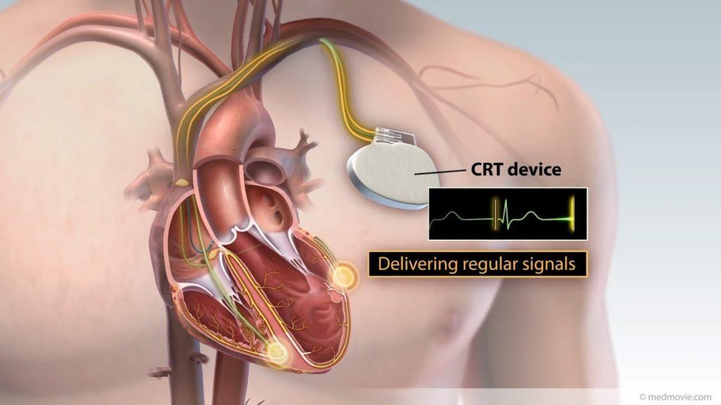
CRT-P – Cardiac Resynchronization Therapy Pacemaker is a specialised pacemaker used to provide cardiac resynchronisation.
In some patients with heart failure, the ventricles do not contract together because of an electrical conduction disorder, most commonly left bundle branch block.
This asynchronous activation can reduce the efficiency of the heart's pumping action.
CRT aims to improve electrical and mechanical coordination between the ventricles.
How does CRT-P work?
A conventional CRT-P system usually has:
- one lead in the right atrium,
- one lead in the right ventricle,
- and an additional lead for the left ventricle.
The left ventricular lead is usually advanced through the coronary sinus, the venous network on the heart's outer surface.
Appropriate programming improves coordination of ventricular contraction.
Who may need CRT?
CRT is mainly used in selected patients with:
- heart failure,
- reduced left ventricular systolic function,
- electrical dyssynchrony on the ECG, particularly left bundle branch block,
- and a specified degree of QRS prolongation.
It may also be considered when a high proportion of ventricular pacing is required and long-term conventional right ventricular pacing could lead to pacing-induced cardiomyopathy, with deterioration in left ventricular systolic function.
In appropriately selected patients, cardiac resynchronisation may:
- improve heart function,
- reduce heart failure symptoms,
- increase functional capacity,
- reduce heart failure admissions,
- and improve prognosis in specific patient groups.
CRT-P or CRT-D?
CRT-P provides pacing and cardiac resynchronisation but does not provide defibrillation or antitachycardia pacing to treat dangerous ventricular arrhythmias.
CRT-D combines cardiac resynchronisation with implantable defibrillator functions.
The choice between CRT-P and CRT-D considers the cause and severity of cardiac dysfunction, risk of serious ventricular arrhythmias, age, other conditions and the overall clinical picture.
What is left bundle branch area pacing – LBBAP?
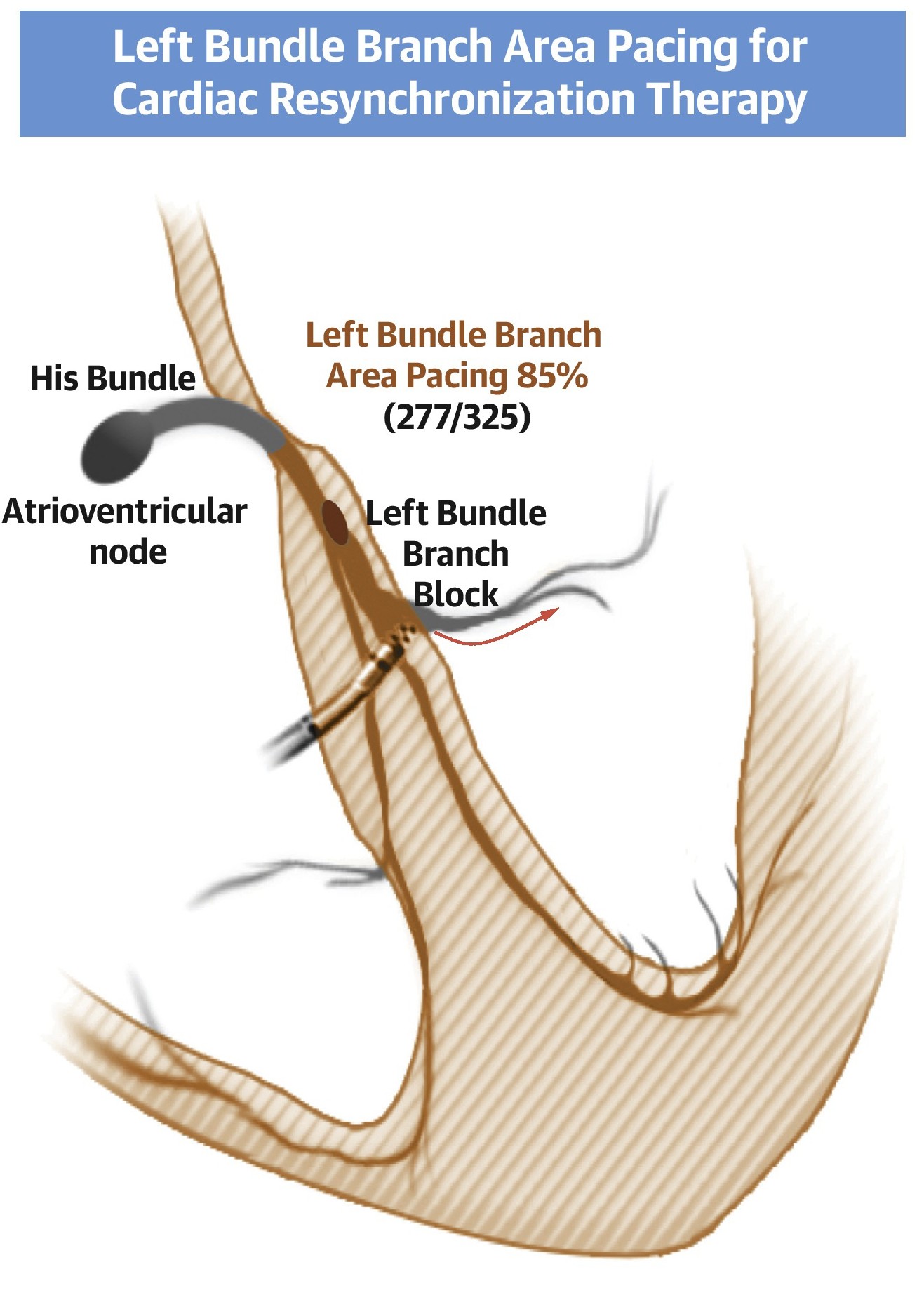
Left Bundle Branch Area Pacing (LBBAP) is a modern form of pacing of the heart's natural conduction system.
Instead of starting an electrical impulse in the right ventricular muscle and allowing it to spread through the myocardium, this technique aims to activate the heart's natural electrical network directly or very close to it.
A specialised lead is advanced and secured deeper within the interventricular septum, near the left bundle branch of the conduction system.
This can achieve more physiological ventricular activation than conventional right ventricular pacing.
Why can this matter?
Long-term pacing exclusively from certain right ventricular sites can produce an abnormal activation sequence in some patients.
When a high proportion of ventricular pacing is expected, preserving more physiological synchrony may be particularly important.
LBBAP may therefore be selected:
- as an alternative to conventional right ventricular pacing,
- when frequent or continuous ventricular pacing is anticipated,
- in selected patients with impaired left ventricular function,
- in some cases requiring cardiac resynchronisation,
- or when conventional CRT lead placement through the coronary sinus is not feasible or does not achieve a satisfactory result.
The best technique differs between patients and is chosen after assessment of the ECG, echocardiogram, underlying conduction disorder and expected pacing requirement.
Before implantation
A full clinical assessment is performed before the procedure.
Depending on the case, this may include:
- an ECG,
- an echocardiogram,
- Holter monitoring or another prolonged rhythm recording,
- blood tests,
- kidney function and electrolyte tests,
- medication review,
- and additional tests when specifically indicated.
Medication
It is particularly important to report all medicines you take, especially:
- anticoagulants,
- antiplatelets,
- diabetes medicines,
- and medicines that affect heart rate.
Do not stop anticoagulants or other cardiac medicines on your own.
You will receive specific instructions about which medicines to continue, adjust or temporarily withhold.
Fasting
Before admission, you will be told how long to fast. Instructions depend on the procedure time and the planned anaesthesia or sedation.
On the day
Before implantation:
- an intravenous line is inserted,
- the necessary cardiac and nursing checks are completed,
- preventive antibiotics are given,
- and the implantation site is prepared under sterile conditions.
How is implantation performed?
Implantation takes place in a specially equipped laboratory with continuous monitoring of heart rhythm, blood pressure and oxygen levels.
Most procedures use local anaesthesia below the collarbone, with light intravenous sedation if needed.
You are usually awake but relaxed and without significant pain.
Creating the pocket
A small incision is made below the collarbone and a pocket is created beneath the skin, or sometimes deeper beneath the muscle, for the generator.
Placing the leads
Leads are advanced through a nearby vein to the heart under X-ray guidance.
Their exact position depends on the system.
In conventional pacing, leads are placed in the right atrium and/or right ventricle.
For LBBAP, the ventricular lead is secured deeper within the interventricular septum to pace the left bundle branch area.
For CRT-P, an additional lead is usually advanced through the coronary sinus into a suitable vein over the left ventricle.
Testing the system
Before completing the procedure, the team carefully checks:
- lead position,
- sensing of the heart's normal electrical signals,
- pacing effectiveness,
- and each lead's electrical parameters.
The leads are connected to the generator and the incision is closed.
Duration varies with the system. CRT implantation or conduction system pacing may take longer than a straightforward pacemaker implant.
What will I feel during the procedure?
After local anaesthetic is given, there should be no significant pain at the incision site.
You may feel:
- pressure or pulling in the area,
- brief discomfort when the local anaesthetic is injected,
- or, less commonly, a momentary heartbeat sensation during lead testing.
The team monitors you continuously and can provide additional pain relief or sedation if needed.
What happens immediately after implantation?
You are transferred to the recovery area or ward for observation.
This usually includes:
- wound checks,
- an ECG,
- full device testing and programming,
- heart rate and blood pressure monitoring,
- and imaging of lead position and the chest when needed.
Minor discomfort or tightness around the wound is expected during the first few days and usually responds to simple pain relief.
Some bruising or mild swelling around the implantation site may occur.
Most patients are able to get up and walk relatively soon after the procedure.
Hospital stay depends on the system type, underlying heart disease, medication and overall condition.
Wound care
Good care of the implantation site supports healing and helps prevent infection.
You will receive specific instructions according to the dressing and wound closure method.
In general:
- keep the wound clean,
- do not remove the dressing early unless instructed,
- do not apply creams, antiseptics or other products without medical advice,
- avoid baths and swimming until the wound has fully healed,
- and check the area daily for infection or a significant haematoma.
A waterproof dressing may allow earlier showering. Always follow your individual discharge instructions.
When should I contact my doctor?
Contact the team promptly if you notice:
- increasing redness around the wound,
- marked or progressive swelling,
- fluid or pus leaking from the wound,
- opening of the wound,
- significant bleeding,
- fever or shivering,
- severe or worsening pain,
- significant swelling of the arm on the implantation side.
Immediate medical assessment is also needed for:
- severe breathlessness,
- chest pain,
- fainting,
- persistent dizziness,
- or prolonged, intense palpitations.
Arm movement and everyday activities
The arm on the pacemaker side should not be kept completely still. Gentle, normal movement helps prevent shoulder stiffness.
During the first weeks, however, you should avoid:
- heavy lifting with that arm,
- sudden or repetitive overhead shoulder movements,
- strenuous upper-body exercise,
- and activities that put pressure or traction on the device area.
Return to work and exercise depends on your occupation, wound healing and underlying heart disease.
Driving
Return to driving depends on:
- the reason for implantation,
- whether fainting occurred beforehand,
- pacemaker dependence,
- whether you hold a private or professional driving licence,
- and applicable legislation.
You will receive individual instructions before discharge.
Pacemaker follow-up
Follow-up is an essential part of treatment.
Before discharge, the device is checked to confirm:
- correct function,
- lead stability,
- adequate battery status,
- and programming suited to your needs.
Regular follow-up is then arranged.
This usually includes:
Wound review
This takes place in the first days or weeks, depending on the closure method and local protocol.
First full device check
Usually performed within 4–6 weeks of implantation.
This assesses:
- the leads,
- pacing thresholds,
- recorded arrhythmia episodes,
- the percentage of pacing,
- battery status,
- and the appropriateness of programming.
Long-term follow-up
After the initial period, checks are performed at intervals determined by the device type and your condition.
Many modern systems also offer remote monitoring, allowing important device information to be transmitted to the medical centre.
Specific follow-up after CRT
For CRT patients, checking that the device functions correctly is only part of follow-up.
Assessment also includes:
- the percentage of effective biventricular or physiological pacing,
- heart failure symptoms,
- functional capacity,
- medical treatment of heart failure,
- and left ventricular function.
Echocardiography may be repeated after an interval to assess the response to resynchronisation.
Some patients need further programming adjustments to achieve the best possible result.
How long does the battery last?
Battery life depends on:
- the device type,
- how often it paces,
- the energy needed for pacing,
- programming,
- and, for CRT, the number of chambers stimulated.
It usually lasts several years.
The battery does not run out suddenly without warning. Regular checks estimate the remaining lifespan well in advance.
When replacement is due, usually only the generator is changed. Existing leads are retained if they work normally.
Are there risks from implantation?
Pacemaker implantation is an established and generally safe procedure. As with any invasive procedure, it carries potential risks.
These include:
- bleeding or a haematoma at the device site,
- wound or system infection,
- lead displacement,
- pneumothorax during vascular access,
- vascular injury,
- perforation of the heart wall and, less commonly, pericardial effusion or tamponade,
- venous thrombosis,
- lead or generator malfunction,
- and, extremely rarely, serious complications requiring further intervention.
Individual risk is affected by age, anticoagulation, kidney function, diabetes, previous devices and overall health.
Specific considerations for CRT-P
Left ventricular lead placement may be technically more demanding because of the coronary venous anatomy.
In some cases, the desired position cannot be reached or phrenic nerve stimulation occurs. This can usually be managed by reprogramming or changing the lead position or settings.
Specific considerations for LBBAP
LBBAP requires precise lead placement deeper within the interventricular septum.
Effectiveness and safety are assessed during implantation using specialised electrophysiological and ECG parameters.
As with any modern technique, selection should reflect anatomy, the underlying condition and expected benefits for the individual patient.
Can I use a mobile phone and electrical appliances?
Most modern pacemakers have a high level of protection against electromagnetic interference.
Everyday household electrical appliances can generally be used normally when working properly.
Mobile phones can also be used, but should not be kept directly over the pacemaker generator for prolonged periods.
Strong electromagnetic fields and certain industrial or medical equipment require special precautions.
Can I have an MRI scan?
Many modern pacing systems are MRI-conditional, allowing MRI under specified conditions.
This depends on:
- the generator,
- the leads,
- the complete implanted system,
- and the conditions under which the scan will be performed.
Before any MRI, inform the device team so compatibility can be confirmed and appropriate programming arranged before and after the scan.
Travel and security checks
After full recovery, most patients with a pacemaker can travel normally.
You will receive a device identification card, which is useful to carry with you.
At airports or other security checkpoints, tell staff you have a pacemaker and follow their instructions.
Will I feel the pacemaker?
The generator may be palpable beneath the skin, particularly in slim people.
After healing, most patients do not feel the pacemaker working and gradually become less aware of its presence in daily life.
Its electrical impulses are extremely small and are not normally felt.
Can I return to normal life?
In most cases, yes.
After healing, most patients can return to everyday activities, work, walking and exercise appropriate to their overall cardiac condition.
A pacemaker is not implanted to restrict activity. In symptomatic bradycardia, treatment can substantially improve dizziness, fatigue, breathlessness and fainting.
Choosing the right technique for you
Modern pacing involves more than deciding whether a patient needs a pacemaker.
Before implantation, it is also important to consider which pacing approach best suits that particular heart.
The choice between:
- conventional pacing,
- left bundle branch area pacing (LBBAP),
- cardiac resynchronisation with CRT-P,
- or a system with defibrillation capability when indicated,
is individualised, aiming both to correct bradycardia and, where possible, preserve or restore the heart's natural synchrony.
The final strategy follows a comprehensive cardiology and electrophysiology assessment and discussion with the patient about expected benefits, alternatives and potential risks.
This page provides general information. The indication for implantation, device type, pacing technique and instructions before and after the procedure are individualised for each patient.